

Skin cancer is the most common type of cancer in the UK, with rates continuing to rise each year. Although types of skin cancer with photos shown online can help you become familiar with their appearance, the different kinds and forms of skin cancer can vary considerably from person to person. Some develop as small, slow-growing patches, while others appear as new or changing moles or sores that do not heal.
Learning to recognise the warning signs can help you seek medical advice sooner, when treatment is often simpler and more effective. However, photographs should only be used as an educational guide. It is not possible to diagnose skin cancer from an image alone, and any suspicious lesion should be assessed by an experienced healthcare professional.
In this guide, we explain the three main types of skin cancer, how they typically appear, and how they differ from one another.
Jump To Section
What Are the Main Types of Skin Cancer?
There are several different forms of skin cancer, but the vast majority of cases fall into three main categories:
| Type of skin cancer | Where it develops | Typical behaviour |
|---|---|---|
| Basal cell carcinoma (BCC) | Basal cells in the deepest layer of the epidermis | Usually grows slowly and rarely spreads to other parts of the body |
| Squamous cell carcinoma (SCC) | Squamous cells in the upper layers of the epidermis | Can grow more quickly and has a greater chance of spreading if left untreated |
| Melanoma | Melanocytes (pigment-producing cells) | Less common but the most serious due to its ability to spread to other organs |
These different kinds of skin cancer each develop from different cells within the skin and often have distinct appearances. Understanding these differences can make it easier to recognise when something doesn’t look quite right.
Basal Cell Carcinoma (BCC)
Basal cell carcinoma is the most common type of skin cancer, accounting for around 80% of non-melanoma skin cancers. It develops from basal cells, which sit at the bottom of the epidermis and continually produce new skin cells throughout life.

The biggest risk factor is long-term exposure to ultraviolet (UV) radiation from the sun or sunbeds. Rather than developing after a single episode of sunburn, basal cell carcinoma usually results from cumulative sun exposure over many years. This is why it most frequently occurs on areas that receive the most sunlight, including the:
- Nose
- Cheeks
- Forehead
- Ears
- Scalp
- Neck
- Shoulders
What Does Basal Cell Carcinoma Look Like?
When looking at types of skin cancer with photos, basal cell carcinoma can appear quite different depending on the individual and the subtype. Some lesions are subtle, while others are more obvious.
Common appearances include:
- A shiny or pearly pink bump
- A small lump with visible blood vessels
- A sore that repeatedly bleeds or crusts before healing and returning
- A flat pink or red patch that slowly enlarges
- A white or scar-like area with poorly defined edges
Unlike many harmless skin blemishes, basal cell carcinomas tend to persist. They often grow slowly over months or years rather than disappearing naturally.
Although basal cell carcinoma rarely spreads elsewhere in the body, it should never be ignored. If left untreated, it can continue to grow into deeper tissues, damaging nearby skin, cartilage and, in some cases, underlying bone. Lesions on the nose, ears and around the eyes are particularly important to assess promptly because treatment can become more complex as they enlarge.

What Will the Photos Typically Show?
Photographs of basal cell carcinoma often demonstrate:
- Smooth, pearly edges
- Pink or flesh-coloured lesions
- Fine surface blood vessels (telangiectasia)
- Central ulceration or crusting
- Slow enlargement over time

Because there are several subtypes of basal cell carcinoma, no single photograph represents every case. Some lesions may resemble eczema, a scar or even a persistent spot, making professional assessment essential whenever a lesion continues to change or fails to heal.
Squamous Cell Carcinoma (SCC)
Squamous cell carcinoma is the second most common form of skin cancer. It develops from squamous cells, which make up most of the outer layer of the epidermis.

Like basal cell carcinoma, it is strongly associated with cumulative sun exposure and is more common in older adults, particularly those with fair skin. However, it can also develop in younger people who have experienced significant UV exposure or have weakened immune systems.
Squamous cell carcinoma most commonly develops on:
- Face
- Ears
- Lips
- Bald scalp
- Back of the hands
- Forearms
- Lower legs
What Does Squamous Cell Carcinoma Look Like?
Among the different forms of skin cancer, squamous cell carcinoma often appears rougher and more inflamed than basal cell carcinoma.
Common features include:
- A firm, red lump
- A rough or scaly patch
- Thickened, crusted skin
- A persistent ulcer
- A lesion that bleeds easily when knocked
Some squamous cell carcinomas grow relatively slowly, while others enlarge over a matter of weeks or months. Unlike basal cell carcinoma, SCC has a greater ability to spread to nearby lymph nodes and other parts of the body if treatment is delayed, although this remains uncommon when diagnosed early.
One important warning sign is a sore that simply refuses to heal. Many people assume these lesions are caused by minor injuries or dry skin, only seeking advice once they continue growing or repeatedly bleed.
What Will the Photos Typically Show?
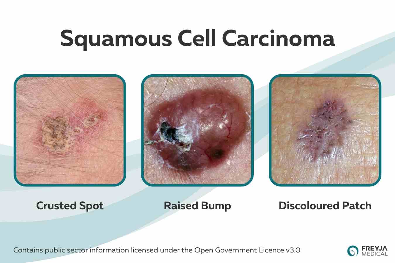
When viewing types of skin cancer with photos, squamous cell carcinoma often appears as:
- Thick, scaly plaques
- Crusted or ulcerated sores
- Raised red nodules
- Rough patches with surrounding inflammation
- Lesions that appear tender or irritated

The appearance can vary depending on its stage and location. Early lesions may closely resemble actinic keratoses or eczema, while more advanced cancers often become thicker, crusted or ulcerated.
Although photographs are useful for recognising general patterns, they should never be used to determine whether a lesion is cancerous. Any skin lesion that changes, bleeds, grows or fails to heal should always be examined by a healthcare professional, who can determine whether further investigation or treatment is needed.
Melanoma
Although melanoma is less common than basal cell carcinoma and squamous cell carcinoma, it is the most serious type of skin cancer because it has a greater ability to spread to other parts of the body if not diagnosed early.
Melanoma develops from melanocytes, the cells that produce melanin, the pigment responsible for giving skin, hair and eyes their colour. It can develop within an existing mole or appear as an entirely new mark on the skin. Unlike basal cell carcinoma and squamous cell carcinoma, melanoma can occur anywhere on the body, including areas that receive little sun exposure, such as the soles of the feet, palms of the hands and underneath the nails.

Early diagnosis is extremely important. When melanoma is identified and treated before it has spread, treatment is often highly successful.
What Does Melanoma Look Like?
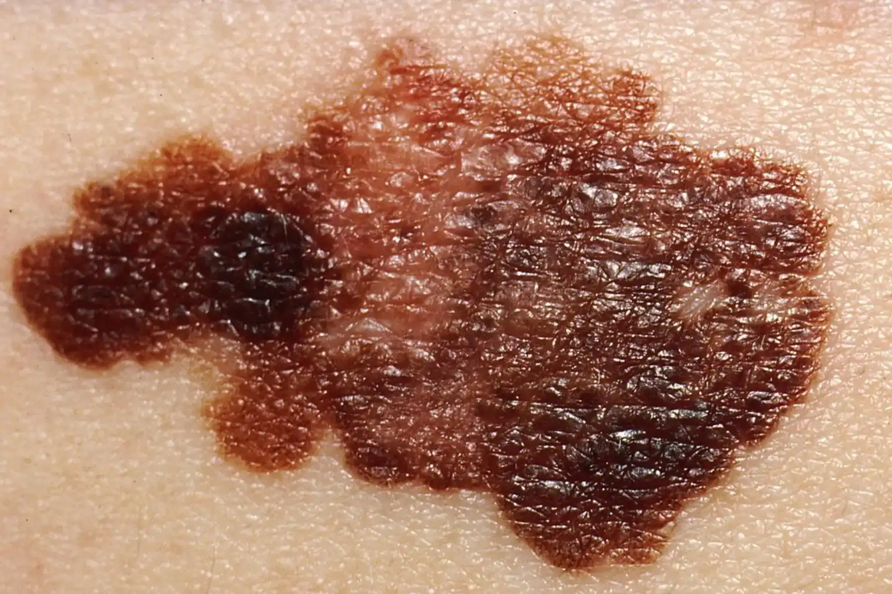
Of all the different kinds of skin cancer, melanoma has the widest variety of appearances. While many melanomas are dark brown or black, they can also be pink, red, blue or even colourless.
A useful way to assess a mole is the ABCDE rule:
- A – Asymmetry: One half does not match the other.
- B – Border: The edges are irregular, blurred or uneven.
- C – Colour: Multiple colours are present within the same lesion.
- D – Diameter: Larger than around 6 mm, although melanomas can be smaller.
- E – Evolving: The mole changes in size, shape, colour or symptoms over time.
You may also notice a mole that begins to itch, bleed, crust or develop a raised area. Any new or changing pigmented lesion should be assessed promptly.

What Will the Photos Typically Show?
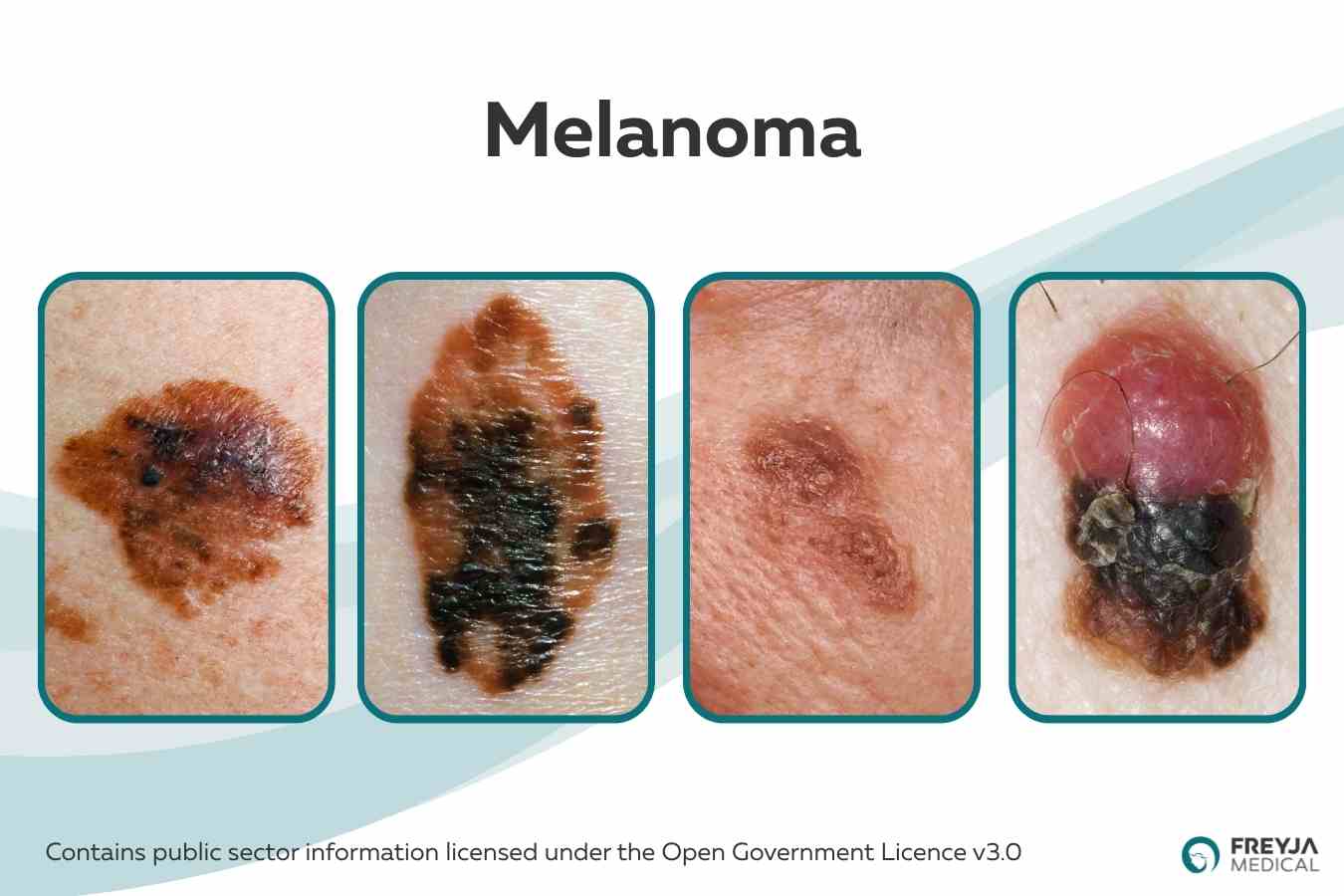
When looking at types of skin cancer with photos, melanoma images commonly demonstrate:
- Irregular or uneven borders
- Variation in colour across the lesion
- An asymmetrical shape
- Enlargement over time
- New or changing moles that stand out from others on the body
However, not every melanoma follows the typical pattern. Some can appear as small pink nodules, while others may develop beneath a fingernail or toenail as a dark streak. This variation highlights why photographs should be used to improve awareness rather than to diagnose skin cancer.
Can Other Skin Conditions Look Like Skin Cancer?
Yes. Many harmless skin conditions can resemble skin cancer, making it difficult to know whether a lesion is something to worry about based on appearance alone.
Some of the most common conditions that can mimic skin cancer include:
- Seborrhoeic keratoses, which are harmless, raised growths that can be brown, black or waxy.
- Actinic keratoses, rough, scaly patches caused by sun damage that can occasionally develop into squamous cell carcinoma.
- Eczema, which may cause persistent red, itchy or flaky patches.
- Psoriasis, producing thick, scaly plaques that sometimes resemble squamous cell carcinoma.
- Benign moles, which may naturally vary in size, colour and shape.
- Cysts, which can appear as smooth, rounded lumps beneath the skin.
Because there is significant overlap in appearance, even experienced clinicians often use a dermatoscope to examine suspicious lesions more closely. In some cases, a biopsy may be recommended to confirm the diagnosis.
If you are ever unsure whether a lesion is harmless, it is always safest to have it assessed professionally rather than relying on online photographs.
When Should You Get a Suspicious Lesion Checked?
While not every skin change is cancerous, certain features should always prompt medical assessment.
Arrange an examination if you notice a lesion that:
- Is new and continues to grow.
- Changes in size, shape or colour.
- Bleeds without obvious injury.
- Crusts repeatedly.
- Does not heal after several weeks.
- Develops persistent itching, tenderness or pain.
- Looks noticeably different from your other moles or skin lesions.
- Changes rapidly over a short period of time.
Regular self-examination can help you become familiar with your skin and identify changes sooner. Pay particular attention to areas that receive frequent sun exposure, including the face, scalp, ears, neck and hands, but remember that skin cancer can develop almost anywhere on the body.
If a healthcare professional suspects skin cancer, they may recommend further assessment, dermoscopy or a biopsy to establish an accurate diagnosis.
How to Book a Consultation
Viewing types of skin cancer with photos can help you become more familiar with how skin cancer may appear, but no photograph can tell you with certainty whether a lesion is cancerous. Basal cell carcinoma, squamous cell carcinoma and melanoma all develop differently, have distinct characteristics and require different approaches to treatment.
Becoming aware of the different forms of skin cancer and the different kinds of skin cancer is an important first step in recognising suspicious skin changes. However, if you notice a mole or lesion that is changing, bleeding, crusting or simply doesn’t look right, the most important action is to seek a professional assessment.
Early diagnosis offers the best opportunity for effective treatment and can help prevent skin cancer from becoming more advanced. If you have any concerns about a suspicious mole or skin lesion, arranging a prompt examination with an experienced dermatology professional can provide reassurance or ensure treatment begins as early as possible.